The projects in our laboratory are multidisciplinar and engage especialists in multiple areas. Here, in concert with computational scientists, clinical surgeons, pathologists and medical oncologists from the Bellvitge University Hospital (HUB), our projects have increased their translational potential while continuing working at a molecular, biochemical and cellular level as well as in vivo by using genetically engineered mouse models.
The Llobet-Navàs laboratory has currently several projects ongoing. For instance, we are embarked in the study of the cellular and physiological mechanisms by which the absence of the miR-424/503 impacts stemness and tumorigenesis in the mammary gland and endometrium. Also, we are exploring the potential role for this miR-cluster in the physiological control of adipose tissue homeostasis. Finally we are also elucidating the role of autophagy in the development and dissemination of endometrial cancer. Scroll down to learn more!
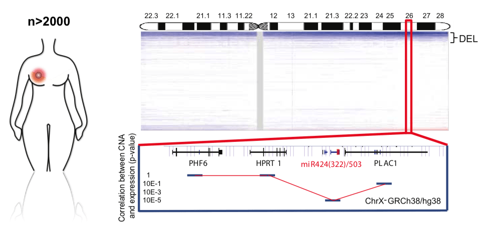
1. miRNAs in breast tumorigenesis and stemness.
Fig.1 - miR-424/503 is lost in breast cancer and is associated with poor prognosis and aggressive subtypes. Copy number profile of the X chromosome in human breast cancers (METABRIC plus TCGA data sets), highlighting the chromosomal region containing the miR-424/503 locus. The correlation between copy number and expression for surrounding genes is included at the bottom.
We have previously identified the miR-424/503 as an important regulator of mammary involution (Llobet-Navas et al. Genes & Development 2014; Llobet-Navas et al. Molecular and Cellular Biology 2014). Mechanistically, we have found that the TGFβ pathway transcriptionally controls its expression in the mammary epithelium, reaching a peak 2-3 days after weaning. By performing in vitro and in vivo functional assays we have discovered that this miRNA unit, by controlling the expression of CDC25A, BCL2 and IGF1R, participates in key signal transduction processes affecting apoptosis and cell cycle. Most recently, our analysis of over 2,000 primary breast cancers has revealed that this miRNA cluster is deleted in about 15% of breast tumors (Rodriguez-Barrueco et al. Genes and Development 2017). In line with these observations, genetically engineered miR-424/503-/- female mice develop mammary tumors after 1 year that are resistant to standard chemotherapy and that the use of IGFR1 and BCL2 inhibitors (already in clinical trials) reinstates treatment response. Following this line of investigation our current and future objectives include, amongst others, the genetic and transcriptomic characterization of breast tumors presenting decreased miR-424/503 expression and the identification of the cell-of-origin that impinges the tumorigenic process.
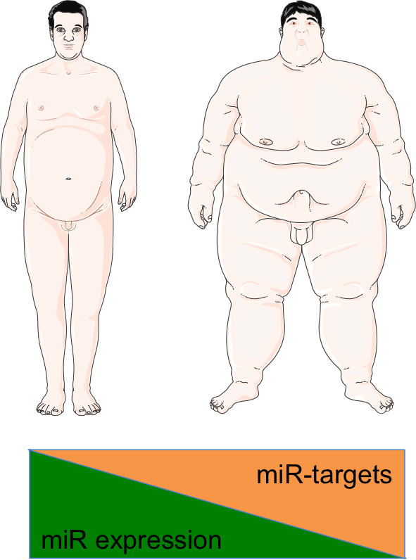
2. miRNAs in adipose tissue.
Fig.2 - Representative illustration of the project.
This line of research that stems from in vivo preliminary data is aimed at deciphering the relevance of the cluster miR-424/503 in the development of hyperplastic adipose tissue (AT). This proposal is structured in eight comprehensive objectives and is designed to dissect the relevance of the cluster miR-424/503 during adipocyte differentiation and adipose tissue development. These objectives will be addressed through the characterization of the miR-424/503-/- obese phenotype, the study of the role of the miR-424/503 in the control of fat cell differentiation and metabolism, the identification of its downstream mRNA regulon and validation of our results using human samples i) by assessing associations between miRNA expression in AT and clinical outputs and ii) by evaluating the impact of polymorphisms affecting the expression of the miR-424/503 cluster.
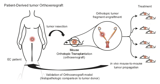
3. Autophagy in the development and progression of endometrial cancer.
Fig.3 - Generation of EC orthoxenografts. Schematic representation of patient-derived orthoxenotransplant implants procedure and treatment used in Eritja et al. Autophagy 2017. EEC tumors were surgically removed and small pieces were implanted in the uterus of recipient female mice. Once engrafted, tumors were propagated to a cohort of 20–45 mice, randomized and treated accordingly.
Endometrial cancer (EC) is the most common gynecological tumor worldwide. Also it is the fourth most common type of tumor in women after breast, colon and lung. EC generally presents a favorable prognosis due to its early detection, when primary surgery treatment is frequently curative, reaching 5-year survival rates over 70%. Despite being treated according to their assessed risk, ~15-20% of these patients continue to relapse after surgery, with recurrence typically affecting to vaginal or pelvic regions, or even presenting metastasis to distant sites hence resulting in poor prognosis. These patients undergo systemic therapy, which may be endocrine therapy or cytotoxic chemotherapy. Unfortunately, these treatments tend to fail and are estimated to only benefit 10–15% of all patients. Current efforts are dedicated to find, in the near future, novel targeted compounds that could be implemented in EC patient management for the first time. In this context we have recently identified autophagy as an essential cellular process endowing endometrial cancer cells with anticancer resistant properties (Eritja et al. Autophagy 2017). In this project we seek to understand and define the genetic triggers and networks involved in the controlled execution of the autophagic process. By utilizing state-of-the-art autophagic flux techniques and high-content fluorescent microscopy devices combined with in vitro EC cell line 3D cultures and PDOs (patient-derived organoids), we expect to unravel novel druggable components of the autophagic machinery. In addition, we will perform in vivo pre-clinical studies by interacting with key collaborators to continue developing and testing our results on an unparalleled and ongoing collection of EC mouse orthoxenotransplants (n>150).
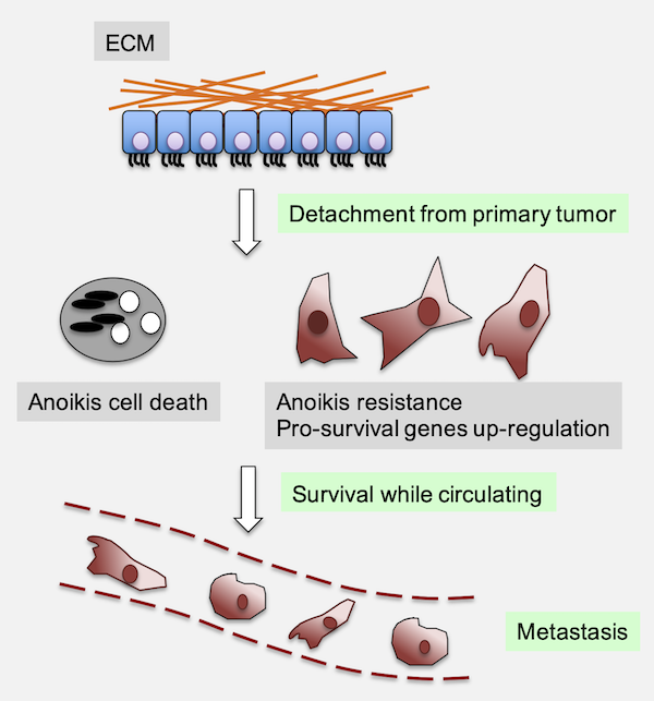
4. Anoikis
Fig.4 - Anoikis evasion contributes to the metastatic process.
The process of anoikis (induced cell death that occurs when the epithelial cell loses its attachment to the extracellular matrix) represents the first line of defense against tumor dissemination, and its evasion represents the initial step for the establishment of future metastasis. The objective of this project is the identification of the key genes involved in anoikis resistance in gynecological tumors, and their exploitation as therapeutic targets. To do this, we combine genome-scale loss-of-function screenings (CRISPRi) with multiomic strategies such as RNA-sequecing or metabolomics, preclinical mouse studies and primary samples.